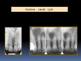
incisive canal radiograph
In addition the angulation of the X-ray beam in panoramic radiography is about 78 from below. Today panoramic radiographs OPGs are routinely used in the dental office for various diagnostic purposes that.

Periapical Radiograph 1 Year After Treatment Bone And Teeth Showing Download Scientific Diagram
The incisive canal also known as the nasopalatine canal is an interosseous conduit through the anterior maxilla connecting the oral and nasal cavities.

. Its radiographic detection remained lower than for the mandibular canal or mental foramen but higher than for the visibility of the lingual foramen. The pear-shaped radiolucency between the apices of the central incisors can be mistaken for periapical pathology or cyst formation. Appearance of the mandibular incisive canal on panoramic radiographs Panoramic radiographs are routinely used in the dental office for various diagnostic purposes.
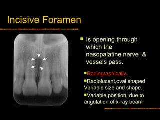
A developmental nonodontogenic cyst that arises from epithelial remnants of the nasopalatine incisive canal adult onset well delineated inverted pear shaped radiolucency interposed between the apices of teeth numbers 8 and 9 root divergence common teeth are vital microscopic. The incisive foramen also known as nasopalatine foramen or anterior palatine foramen is the oral opening of the nasopalatine canal. The mean endpoint was approximately 1098 and 1026 mm anterior to the mental foramen for left and right side respectively without a.
In the study by Jacobs et al 1 on 230 spiral CT scans of the mandible the incisive canal could be identified in 93 of the spiral CT scans. The presence of the cyst is presumed if the width of the foramen exceeds 1 cm or if enlargement can be demon-strated on successive radiographs. Mean canal length was 1863 235 mm and males have significantly longer incisive canal than females.
Other shapes on radiographic imaging include cylindrical banana spindle or hourglass. This might be explained by the fact that the incisive canal is less corticalized and has a smaller diameter than the mandibular canal. Sse and respiratory epithelium with neurovascular bundle.
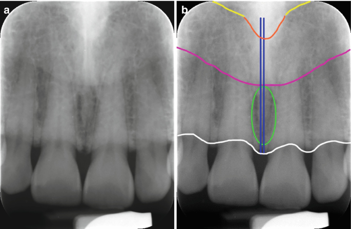
It is located in the maxilla in the incisive fossa midline in the palate posterior to the central incisors at the junction of the medial palatine and incisive sutures. The incisive canal is located in the anterior part of the hard pa late and serves as a communication between the oral and nasal cavities. Within this canal lies the nasopalatine nerve and the vascular anastomosis between the greater palatine and sphenopalatine arteries.
This results in some distortion of the actual mandibular anatomy and may lead to misinterpretation. This study aimed to evaluate the visibility of neurovascular structures in the. Its appearance is quite variable due to normal anatomic variation and due to the operators angulation of the x-ray beam.
Several authors have reported different dimensions of radiolucency as diagnostic of. The incisive foramen is important because it is a potential site of cyst formation. On imaging the shape of the canal has been observed most commonly to be a funnel or Y-shape 14.
The nasopalatine duct cyst occurs in the nasopalatine or incisive canal and it may be difficult to decide on a radiograph whether radiolucency in that area is a cyst or a large incisive foramen. A well-defined incisive canal could be detected in the majority of spiral CT scans. On periapical x-ray images the incisive foramen is located in the midline between the roots of the central incisors.
Knowledge of the anatomy in the region between the mental foramens is still poorly documented although correct identification of the anatomical structures in this region is important for the success of surgical procedures In the literature complications can be found due to anatomical variation in the inferior alveolar nerve because this nerve can. Popularly known as nasopalatine canal is a radiolucent tube shaped area located in between the maxillary central incisors. In some radiographsB the incisive canal Fig.
The embryology of the canal has led to interesting theories explaining. Only in a very few radiographs will the incisive canal or nasopalatine canal be. Coronoid process is the thin triangular-shaped process of the anterosuperior aspect of the ramus.
Results The incisive canal was found in 87 of the scans. The incisive foramen generally appears in most panoramic radiographs though not with the clarity seen in periapical radiographs. Usually only the inferior border of the orbit is visible over the panoramic radiograph Incisive canal.
It can be single or multiple. 3B can be seen leading to the incisive fora-. Radiological imaging methods evaluating the incisive canal further corroborate morphological variances in shape course angulation and direction.
Assessments included 1 mesiodistal diameter 2 labiopalatal diameter 3 length of the incisive canal 4 shape of incisive canal and 5 width of the bone anterior to the incisive foramen.

The Radiology Of Developmental Dental Defects Demystified An E Based Learning System Intechopen
Normal Radiographic Anatomical Landmarks

Anterior Loop Of The Mental Nerve And Its Radiologic Imaging A Review Semantic Scholar

Radiographic X Ray Taken After Root Canal Treatment On Tooth 12 A Download Scientific Diagram

Pdf Visibility Of Maxillary And Mandibular Anatomical Landmarks In Digital Panoramic Radiographs A Retrospective Study Semantic Scholar

Maxillary Anterior Landmarks Intraoral Radiographic Anatomy Continuing Education Course Dentalcare Com
Route Of The Incisive Canal Of The Mandible Mic Download Scientific Diagram
Intra Oral Radiographic Anatomical Landmarks

Xmlinkhub

Normal Radiographic Anatomical Landmarks

Cone Beam Ct Images Showing A B Sagittal Cuts Of A Bifid Incisive Download Scientific Diagram

A Panoramic Radiograph Shows The Anterior Loop And Incisive Canal Download Scientific Diagram
Normal Anatomical Landmarks In Dental X Rays And Cbct Springerlink

Pdf The Evaluation Of Visibility Of Mandibular Anatomic Landmarks Using Panoramic Radiography Semantic Scholar

Intraoral Radiographs Identifying Normal Anatomy Today S Veterinary Practice

6 Essentials Of Dental Radiographic Analysis And Interpretation Pocket Dentistry

6 Essentials Of Dental Radiographic Analysis And Interpretation Pocket Dentistry

Mouth Incisive Canal Cyst Professional Radiology Outcomes

Opg Showing Incisive Foramen And Mental Foramen Download Scientific Diagram


Comments
Post a Comment